


Ultrasonography in Obstetrics and Gynecology

by Premier Hospitals | July 24, 2019 |
No doctor can see inside of your body. Then how do they know the changes or causes in your body? You are thinking is right! The answer is ultrasonography. Commonly known as a scan. Ultrasonography is a diagnostic medical procedure in which the high-frequency sound waves are transmitted into body deeper organs and bounce-off. This procedure is helping your radiologist to view or picturise, and diagnose the conditions of your inner organs, vessels and tissues without an incision. It helps in analysing the growth of the foetus and deformities if any. This article is all about what is Ultrasonography, its benefits, how it works and how it helps in diagnosis. Continue your reading now! The introduction of ultrasound in gynaecology and obstetrics has a significant impact on patient care because it allows better visualization of the fetus and placenta in obstetrics and visualisation of maternal organs in gynaecology. It is important to mention here that such visualisation has high video clarity paving the way to advanced diagnosis and appropriate treatment. Understanding the physical basis of ultrasound is very important, and also doctor should have a fundamental knowledge of operating ultrasound instrument, and understand the safety and bioeffects of this technology. This chapter introduces a detailed description of ultrasonography, its uses, procedure, by whom it is operated, and uses in obstetrics and gynaecology. What is an Ultrasonography?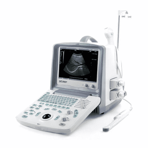 Ultrasound sonography uses high-frequency sound waves (ultrasonic waves) to produce images of internal organs and other tissues. Ultrasound is painless, relatively inexpensive, and is considered very safe during pregnancy.
Ultrasound images help diagnose various diseases and conditions. The sonographic idea comes from sonar technology, which uses sound waves to detect underwater objects.
How does it work?
Ultrasound sonography uses high-frequency sound waves (ultrasonic waves) to produce images of internal organs and other tissues. Ultrasound is painless, relatively inexpensive, and is considered very safe during pregnancy.
Ultrasound images help diagnose various diseases and conditions. The sonographic idea comes from sonar technology, which uses sound waves to detect underwater objects.
How does it work?
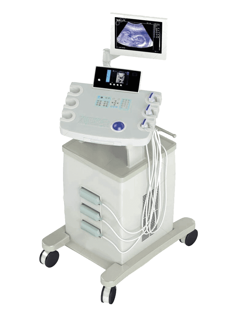 Devices called transducers to convert electrical currents into sound waves that are sent to body tissues. Sound waves bounce off the structure in the body and are thrown back into the transducer, which converts waves into electrical signals. Computers changed the pattern of electrical signals into images displayed on a monitor and recorded on film, on videotape, or as digital computer images. X-rays are not used.
Ultrasonography cannot be used to visualize bones because it is too dense to penetrate. Also, the intestinal tract and normal lung tissue are not easily identified by ultrasound because air or gas prevents the formation of ultrasound images. Ultrasonography can be used with other diagnostic procedures or alone.
When does the
By Whom and to Whom Ultrasonography is Done.
A skilled ultrasound technologist can use sonography to answer questions in the body that can be asked by the doctor. As a rule, radiologists monitor ultrasound tests and report the results. However, other types of doctors can also use ultrasound as a diagnostic tool. For example, obstetricians use ultrasound to examine the fetus during pregnancy. Surgeons and emergency doctors use ultrasound in bed to assess abdominal pain or other problems.
Ultrasound helps in creating images of the soft tissue structures such as the gallbladder, liver, heart, kidney, female reproductive organs, and even babies still in the womb. Ultrasonography can also detect blockages in blood vessels.
If a patient visits the hospital with any type of problem in a reproductive organ or about the problem related to pregnancy, then the ultrasound is suggested to see an internal problem. Ultrasound is a medical device which detects and evaluates, diagnoses, and helps the doctor to cure diseases all over the body, especially in obstetrics and gynaecology.
Procedure to Use Ultrasonography in Gynecology
If your stomach is being examined, you may be asked not to eat and drink for several hours before the test.
As a rule, the examiner applies the thick gel to the site to be examined to ensure good sound transmission. A manual transducer is placed on the skin and transferred to the area to be evaluated.
To assess gynaecology reproductive parts of the body, the examiner inserts a transducer into the body, for example, into the vagina to image the uterus and ovary, or into the anus to visualize the prostate.
Biometric measurement of the uterus includes its length, height, and width, and the endometrial thickness measured in sagittal plane.and each ovaries height and width is measured, and evaluate for the presence of fluid and other abnormalities. After the test, most people can immediately resume their usual activities.
Procedure to use Ultrasonography in Obstetrics
As a rule, the examiner applies the thick gel to the site to be examined to ensure good sound transmission. A manual transducer is placed on the skin and transferred to the area to be evaluated.
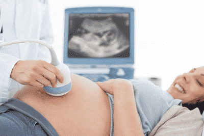
Traditional obstetrical sonograms are performed by attaching sensors to the abdomen of a pregnant woman. One variant, transvaginal sonography, is performed with a probe in the woman's vagina. With Doppler sonography, the fetal heart and blood vessels can be examined for signs of abnormalities.
Uses of Ultrasonography
We at Premier Hospital uses ultrasound in obstetrics and gynecology for the following:
Uses of Ultrasonography in Obstetrics
Devices called transducers to convert electrical currents into sound waves that are sent to body tissues. Sound waves bounce off the structure in the body and are thrown back into the transducer, which converts waves into electrical signals. Computers changed the pattern of electrical signals into images displayed on a monitor and recorded on film, on videotape, or as digital computer images. X-rays are not used.
Ultrasonography cannot be used to visualize bones because it is too dense to penetrate. Also, the intestinal tract and normal lung tissue are not easily identified by ultrasound because air or gas prevents the formation of ultrasound images. Ultrasonography can be used with other diagnostic procedures or alone.
When does the
By Whom and to Whom Ultrasonography is Done.
A skilled ultrasound technologist can use sonography to answer questions in the body that can be asked by the doctor. As a rule, radiologists monitor ultrasound tests and report the results. However, other types of doctors can also use ultrasound as a diagnostic tool. For example, obstetricians use ultrasound to examine the fetus during pregnancy. Surgeons and emergency doctors use ultrasound in bed to assess abdominal pain or other problems.
Ultrasound helps in creating images of the soft tissue structures such as the gallbladder, liver, heart, kidney, female reproductive organs, and even babies still in the womb. Ultrasonography can also detect blockages in blood vessels.
If a patient visits the hospital with any type of problem in a reproductive organ or about the problem related to pregnancy, then the ultrasound is suggested to see an internal problem. Ultrasound is a medical device which detects and evaluates, diagnoses, and helps the doctor to cure diseases all over the body, especially in obstetrics and gynaecology.
Procedure to Use Ultrasonography in Gynecology
If your stomach is being examined, you may be asked not to eat and drink for several hours before the test.
As a rule, the examiner applies the thick gel to the site to be examined to ensure good sound transmission. A manual transducer is placed on the skin and transferred to the area to be evaluated.
To assess gynaecology reproductive parts of the body, the examiner inserts a transducer into the body, for example, into the vagina to image the uterus and ovary, or into the anus to visualize the prostate.
Biometric measurement of the uterus includes its length, height, and width, and the endometrial thickness measured in sagittal plane.and each ovaries height and width is measured, and evaluate for the presence of fluid and other abnormalities. After the test, most people can immediately resume their usual activities.
Procedure to use Ultrasonography in Obstetrics
As a rule, the examiner applies the thick gel to the site to be examined to ensure good sound transmission. A manual transducer is placed on the skin and transferred to the area to be evaluated.
Traditional obstetrical sonograms are performed by attaching sensors to the abdomen of a pregnant woman. One variant, transvaginal sonography, is performed with a probe in the woman's vagina. With Doppler sonography, the fetal heart and blood vessels can be examined for signs of abnormalities.
Uses of Ultrasonography
We at Premier Hospital uses ultrasound in obstetrics and gynecology for the following:
Uses of Ultrasonography in Obstetrics
- Prenatal genetics and diagnostics
- Fetal aneuploidy ultrasound examination in the first and second trimesters
- Ultrasonography of the initial first trimester
- Assessment of fetal anatomy in the first trimester
- Biometry and fetal growth
- Multiple pregnancy ultrasound examinations
- Ultrasound examination for normal fetal anatomy
- Ultrasound evaluation is done to see the fetal central nervous system
- Ultrasound examination of the face and neck of the fetus
- Fetal musculoskeletal system
- Ultrasound examination of Fetal thorax
- Fetal heart ultrasound examination
- Ultrasound examination of the fetal digestive tract and abdominal wall
- Fetal genitourinary tract
- Ultrasound properties of the fetal syndrome
- Ultrasound Evaluation of Hydrops Fetalis
- Ultrasound Evaluation of the Gravid Cervix
- Ultrasound examination of the placenta, membrane, and umbilical cord
- Amniotic fluid volume in fetal health and disease
- Antepartum Fetal Surveillance and the role of ultrasound
- Role of Magnetic Resonance Imaging in Obstetrics
- Role of Sonography in Fetal Procedures
- The role of sonography in fetal procedures
- Obstetric Ultrasound Imaging and the Obese Patient
- Endometrial studies to assess the suitability of ET techniques in vitro fertilization

- Normal pelvic anatomy and transvaginal sonography
- Abnormal uterine bleeding, the role of ultrasonography
- Uterine ultrasound examination
- Assessment of pelvic pain in reproductive age
- Ovarian ultrasound examination
- Ultrasound examination of the fallopian tube
- Assisted infertility and reproductive sonography
- Ectopic pregnancy
- Gynecological sonography in children and adolescents
- Ultrasonography and Magnetic Resonance Imaging in Urogynaecology
- Uses in the assessment of gynecological diseases
- Sonographic role in gynecological procedures