


How Is A Heart Condition Diagnosed?
by Premier Hospitals | May 1, 2020 |
Disorders of the heart are referred to as Heart Disease or Cardiovascular diseases. They include narrowed or blocked blood vessels that can cause heart attacks, chest pain (angina pectoris), or stroke and also other heart conditions, such as those that affect the heart muscle, valves or rhythm. Many forms of heart disease can be prevented or treated if diagnosed early. The approach to a diagnosis is as follows: Your doctor first records your medical and personal history, checks your family history, medical records and previous symptoms, and laboratory tests and electrocardiograms. He/She will then decide to do additional tests if necessary. For the diagnosis of Heart disease, there are various tests that can be used. These tests can be non-invasive (which means no instruments are inserted into the body) or invasive (require tools to be inserted into the body). The tests ordered can vary greatly depending on the type of heart disease. Different types of a diagnostic test for heart conditions: 1. Check your heart rate.

- Complete blood count (CBC): CBC is a series of tests that measure your red blood cells, white blood cells and platelets.
- Sodium and potassium levels: Sodium and potassium levels are measured to determine problems with electrolytes in body fluids.
- Blood urea nitrogen and creatinine: Blood urea nitrogen and creatinine are measured to check kidney function.
- Fasting glucose: Fasting glucose is done to diagnose diabetes or prediabetes.
- ALT and AST: ALT and AST are performed to detect inflammation or liver damage.
- TSH: TSH is measured to check thyroid function.
- Electrocardiogram (ECG): This test is a general test that records the electrical activity of the heart. It helps to identify heart attacks, abnormal rhythms (arrhythmias) and can also detect damage to the heart muscle.
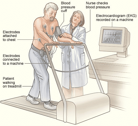
- Stress test: This is also known as a treadmill or ECG exercise. This test is done to monitor your heart when you run on a treadmill. Your doctor will also monitor your breathing and blood pressure. Stress tests can be used to identify coronary artery disease or to determine a safe level of exercise after a heart attack or heart surgery. This test can also be done with the help of particular drugs that similarly stress the heart as physical exercise. Sometimes stress tests collect ECG information along with images of ultrasound of the heart. It is known as exercise or stress echocardiogram. It is more sensitive and specific than just an ECG stress test.
- Transthoracic echocardiogram (echo or TTE): Echo is a non-invasive test that uses sound waves to assess the chambers and valves of your heart and how well it pumps. The echo sound waves create a real-time image on the monitor with the ultrasound probe which is transmitted through the skin from your heart.
- Transesophageal Echocardiogram (TEE): This test resembles transthoracic echocardiogram. But you are taken with medication to help you stay sedated. It is more invasive because the probe is inserted into your body. In this test, you swallow a small probe the size of your thumb. The probe runs down the oesophagus just behind the heart. It allows a closer look at the structure and function of the heart than a standard echocardiogram. It is a better test to check the structure and function of the heart valve. Your doctor may be able to recognize blood clots in the heart better.
- Positron emission tomography (PET): This is a nuclear scan that provides information about blood flow through the coronary arteries to the heart muscle:
- PET F-18 FDG scan (fluorodeoxyglucose): This particular PET scan uses a form of radioactive glucose to determine whether particular areas of heart tissue are permanently damaged. Your doctor can use it after a heart attack to determine which procedures, such as angioplasty, stenting, or bypass surgery, can help. Test technicians inject glucose solutions into your bloodstream through IV. Then a special camera takes a photo when the solution collects in your heart.
- Thallium scanning or myocardial perfusion: It involves IV injection (radioactive tracer) and a special camera like pet scanning:
- Resting SPECT thallium scan or myocardial perfusion scan: Nuclear scanning is done while you are resting. Areas of the heart muscle that do not receive blood flow at rest are examined.Â
- Myocardial perfusion: Nuclear scanning is carried out during exercise. The examination is performed on areas of the heart muscle that do not receive enough blood during activity.
- Adenosine or persantine thallium scan or myocardial perfusion: Nuclear scanning is done when you can't exercise. The examination is performed on areas of the heart muscle that do not receive enough blood. It uses special drugs that stress the heart, as a similar way as exercise does.
- MUGA scan/radionuclide angiography (RNA): Like PET scanning, this test involves IV injection of a radioactive tracer and a special camera:
- RGBPS (Resting gated blood pool scan, resting MUGA, radionuclide angiography at rest: Nuclear scan to evaluate how well the heart wall moves and how much blood pumps during each heartbeat when you rest.
- EGBPS (exercise gated blood pool scan), MUGA exercises, or radionuclide angiography while exercising: Nuclear scans to assess how well the heart wall moves and how much blood is pumped with each heartbeat as soon as you walk on a treadmill.
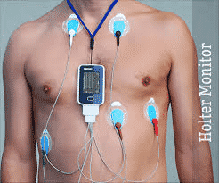
- Event recorder: For this test, you wear a small, portable, battery-powered device that has been used for ECG recording for several weeks. Each time you experience symptoms, press the recorder button to record ECG samples. You will send this sample to the doctor's office for assessment as soon as possible. Other types of event recorders do not use cable. Instead, a small electrode card is placed on the skin above the heart.
- Tilt test: Your doctor connects you to an ECG and blood pressure monitor. You are fixed to a table that tilts you from lying to upright. You can use this test to determine whether a sudden drop in blood pressure (orthostatic hypotension) occurs when standing. You may need this test if you have frequent fainting spells.
- Electrophysiological examination: For this test, an isolated electric catheter is inserted through a large vein in the thigh into the heart. It is used to test the heart's electrical system. It will help your doctor find out what can cause irregular heartbeats.
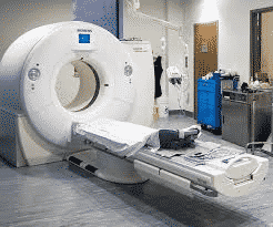
- MRI of the heart: This procedure uses a combination of large magnets, radio frequencies, and computers to take detailed pictures of organs and structures in your body. Your doctor can order a heart MRI to check the heart valves and major blood vessels. It can also detect coronary artery disease and how much damage it causes. It can also assess heart problems that have occurred since birth. It can detect tumours and other issues. A doctor can order this test before other procedures such as angioplasty or coronary artery stenting as well as heart or blood vessel surgery:
- Computer heart tomography: This test is similar to an MRI but a CT scanner is used instead. In cardiac tomography, you lie on a table with a doughnut-shaped machine. An x-ray inside the device rotates around the body and collects images of heart and chest.